Dr Balvinder HandaPerth Cardiovascular Institute Refer a patient

GP Handbook · Practical Quick Reference

Common arrhythmias for general practice

A practical handbook for GPs managing palpitations, atrial fibrillation, SVT, ventricular ectopy, bradyarrhythmias and unexplained syncope — with a clear pathway from recognition and initial investigation to electrophysiology referral.

Dr Balvinder Handa MBBS BSc MRCP FRACP PhD Consultant Cardiologist & Cardiac Electrophysiologist
Perth Cardiovascular Institute Mounts Bay Road · Hollywood / Nedlands · St James · Midland
Public appointment Sir Charles Gairdner Hospital
AF / Flutter / AT SVT / WPW Ventricular ectopy / NSVT Bradycardia / Devices

Perth Cardiovascular Institute

Dr Balvinder Handa

Consultant Cardiologist / Cardiac Electrophysiologist

Arrhythmias · Ablation · Devices · Inherited Cardiac Conditions
Arrhythmia Care Delivered Across Private and Public Hospitals in PerthCardiac electrophysiology, ablation and device care through Perth Cardiovascular Institute, with a public appointment at Sir Charles Gairdner Hospital.

GP quick referral guide

When should an arrhythmia patient be referred?

Use the rhythm, symptoms and underlying substrate to decide urgency. These are practical prompts rather than a substitute for clinical judgement or local emergency pathways.

Emergency / ED now

Do not wait for an outpatient review

  • Ongoing haemodynamic instability with a tachyarrhythmia or bradyarrhythmia
  • Sustained broad-complex tachycardia / suspected VT or VF
  • Syncope with persistent high-grade AV block or severe symptomatic bradycardia
  • Cardiac arrest, recurrent syncope with ongoing arrhythmia, or acute heart failure related to the rhythm
Prompt EP / cardiology referral

Arrhythmia likely to need specialist management

  • WPW / ventricular pre-excitation on ECG, particularly with palpitations or AF
  • Recurrent abrupt-onset / abrupt-offset SVT
  • Unexplained syncope with conduction disease, an abnormal ECG or injury
  • NSVT or ventricular ectopy with symptoms, abnormal ECG/echo, high burden or concerning family history
  • Suspected long QT syndrome, Brugada syndrome or inherited arrhythmia
Early / routine referral

Useful when treatment choices need discussion

  • Symptomatic AF, atrial flutter or atrial tachycardia
  • Younger AF, recurrent AF, drug intolerance or a rhythm-control / ablation discussion
  • Palpitations captured on ECG or ambulatory monitoring where the diagnosis or significance is uncertain
  • Bradycardia, pauses or chronotropic incompetence associated with symptoms
  • Cardiac device assessment or follow-up

What helps with a referral

Attach what is already available

A referral does not need every investigation completed first. For urgent problems, refer or escalate without delaying care.

  • 12-lead ECG — during symptoms if available
  • Holter / event monitor report and representative traces
  • Echocardiogram if already performed
  • Current medications, including anticoagulation and anti-arrhythmics
  • Symptom pattern: abrupt vs gradual onset, duration, exercise relationship and syncope
  • Relevant bloods: electrolytes, thyroid function and haemoglobin where appropriate
  • Family history of sudden death, cardiomyopathy or inherited arrhythmia

01 · General approach

Arrhythmia in general practice

Most rhythm presentations become easier once you answer 4 questions: what rhythm is this, is there structural heart disease, is there a reversible trigger, and does the patient need specialist treatment?

ECG

12-lead ECG

Look for the arrhythmia itself, conduction disease, pre-excitation, channelopathy patterns and QT abnormalities.

MON

Ambulatory monitoring

Holter monitoring and single-lead ECG devices help capture intermittent palpitations and quantify ectopic burden.

ECHO

Structural assessment

Use echocardiography first line; cardiac MRI becomes important where cardiomyopathy, scar or ventricular arrhythmia is suspected.

LAB

Reversible causes

Check thyroid function, electrolytes and other relevant secondary triggers such as anaemia, alcohol excess and uncontrolled hypertension.

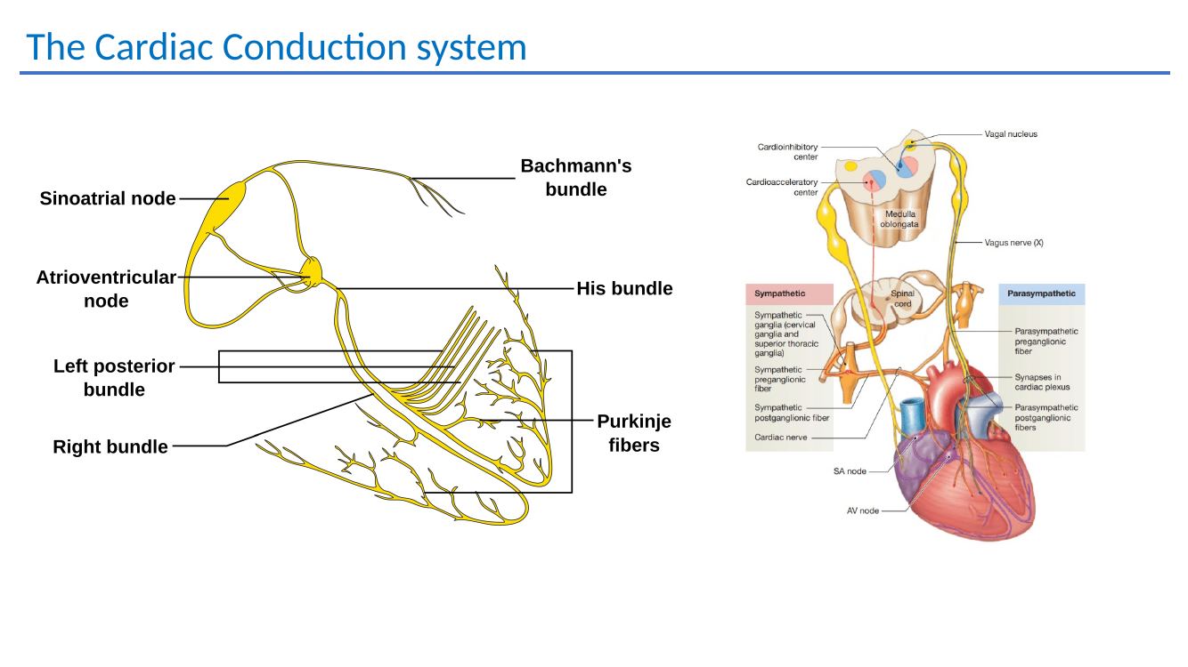
Cardiac conduction system teaching diagram
The cardiac conduction system — useful context when separating atrial tachyarrhythmias, AV nodal re-entry and conduction disease.

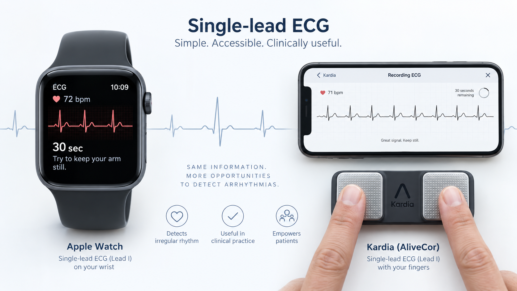
Single-lead ECG devices

One of the most useful tools for intermittent palpitations

For patients whose symptoms come and go, a single-lead ECG recorded during the episode can be far more useful than a normal ECG taken later in the clinic. Apple Watch ECG and AliveCor / Kardia devices are common examples, and patients can usually save or export the tracing for review.

Practical GP message

Ask the patient to record the rhythm while they are symptomatic.

The value is the ECG trace itself. It can provide symptom–rhythm correlation, document AF, show a regular tachycardia during suspected SVT, demonstrate ectopy or bradycardia, and often shorten the pathway to a definitive diagnosis.

1Sit still and record as soon as symptoms start.
2Aim for at least 30 seconds of clean tracing where possible.
3Label the recording with symptoms, time and activity.
4Save/export the PDF or tracing and attach it to the referral.
Apple Watch and Kardia single-lead ECG devices with ECG trace examples
Single-lead ECG devices can be very helpful for intermittent symptoms. Apple Watch and Kardia-type devices allow patients to capture a rhythm strip at the time of palpitations, which can then be reviewed clinically or attached to the referral.
WATCH

Apple Watch ECG

Single-lead, Lead-I-like recording

  • Excellent because the device is already on the patient's wrist when symptoms occur.
  • Produces a short ECG that can be reviewed on the iPhone and exported or shared.
  • Particularly useful for documenting AF, regular tachycardia, ectopy and bradycardia during symptoms.
  • The ECG app is separate from passive irregular-rhythm notifications; a notification alone is not the same as an interpretable ECG tracing.
Apple ECG information
KARDIA

Kardia / AliveCor

Handheld single-lead ECG; 6-lead versions also available

  • A patient places fingers on the electrodes and records a 30-second rhythm strip using a phone or tablet.
  • Simple to keep at home or carry for recurrent episodes that are difficult to capture on a Holter.
  • The app may classify recordings as normal, possible AF, bradycardia, tachycardia or unclassified, depending on device/software.
  • The stored ECG can be sent directly to the GP or included with an electrophysiology referral.
AliveCor device information
GP

How I use these recordings

The tracing is more important than the automated interpretation

  • AF: a clear 30-second single-lead ECG can provide diagnostic rhythm documentation when interpreted clinically.
  • SVT: a regular narrow-complex tachycardia captured during symptoms is extremely useful, even if a single lead cannot always define the exact SVT mechanism.
  • Ectopy: recordings can confirm that the patient's “skipped beats” correspond to PACs or PVCs.
  • Bradycardia: a recording made during dizziness can reveal marked sinus bradycardia or pauses, although intermittent conduction disease may still require longer monitoring.
Important limitation

A single lead is not a replacement for a 12-lead ECG.

It is less useful for defining QRS morphology, localising ventricular ectopy or an accessory pathway, assessing QT, ischaemia or conduction patterns, and distinguishing every tachycardia mechanism. Automated device classifications can also be wrong — review the actual trace.

Do not delay urgent care to obtain a wearable ECG. Syncope, ongoing chest pain, haemodynamic compromise, severe dyspnoea or sustained broad-complex tachycardia should follow the appropriate emergency pathway.
Useful referral attachmentIf your patient has captured an arrhythmia on Apple Watch, Kardia or another ECG-capable device, attach the ECG PDF or screenshot with the referral. A good symptom–rhythm recording can materially accelerate the diagnosis.
Refer to Dr Handa →

02 · Atrial fibrillation

AF: recognise it, assess stroke risk, then choose rate or rhythm control

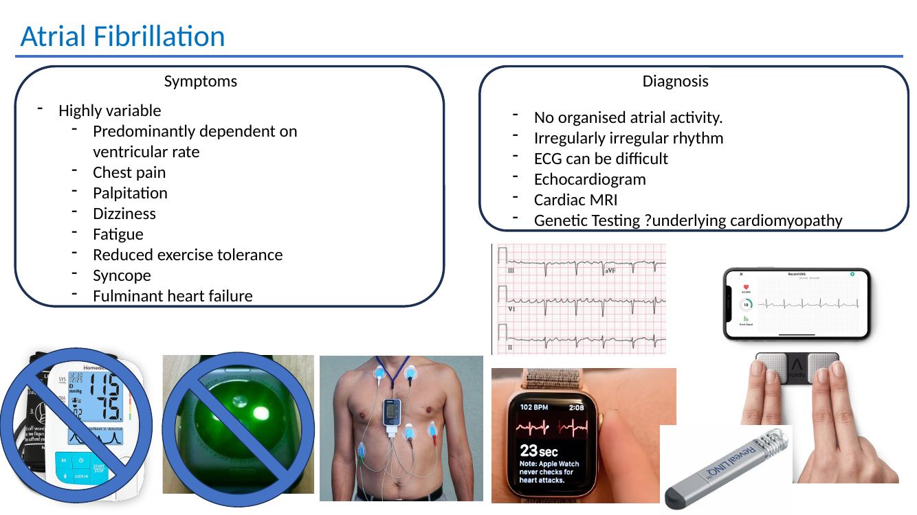
Symptoms are highly variable and often depend on the ventricular rate. Typical features include palpitations, chest pain, dizziness, fatigue, reduced exercise tolerance, syncope and, occasionally, acute heart failure.

Diagnosis

  • No organised atrial activity
  • Irregularly irregular ventricular rhythm
  • ECG diagnosis can occasionally be difficult because of artefact or organised-looking fibrillatory activity
  • Assess for structural disease with echocardiography; consider CMR/genetic evaluation where an underlying cardiomyopathy is suspected
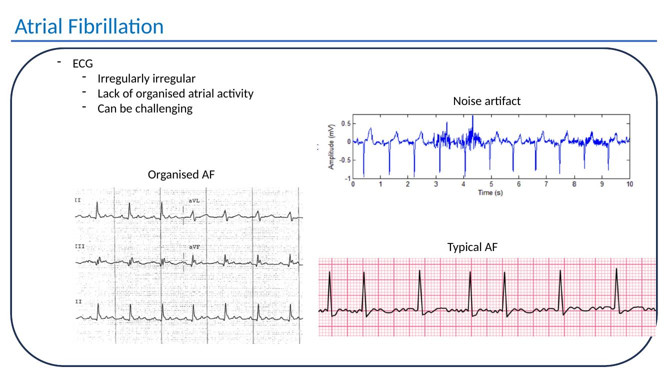
Atrial fibrillation ECG examples
AF ECG examples from the teaching deck, including typical AF and mimics/artefact.
GP practice point: confirm AF on ECG, assess stroke prevention separately from symptom control, and address modifiable cardiovascular risk factors and comorbidities. A younger or symptomatic patient often benefits from an earlier rhythm-control discussion rather than simply escalating rate-control therapy.

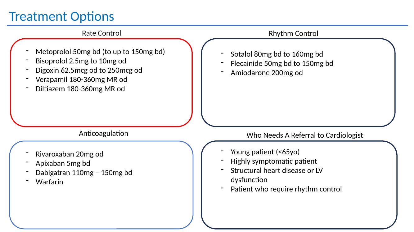
Drug options used in the presentation

Rate control

AV nodal blockade

  • Metoprolol 50 mg bd, titrating as required
  • Bisoprolol 2.5–10 mg od
  • Digoxin 62.5–250 mcg od
  • Verapamil MR 180–360 mg od
  • Diltiazem MR 180–360 mg od
Rhythm control

Anti-arrhythmic therapy

  • Sotalol 80–160 mg bd
  • Flecainide 50–150 mg bd
  • Amiodarone 200 mg od

Choice depends on age, structural substrate and contraindications.

Anticoagulation

Stroke prevention

  • Rivaroxaban 20 mg od
  • Apixaban 5 mg bd
  • Dabigatran 110–150 mg bd
  • Warfarin

Use according to standard stroke-risk assessment and renal/bleeding considerations.

Catheter ablation

A definitive rhythm-control option

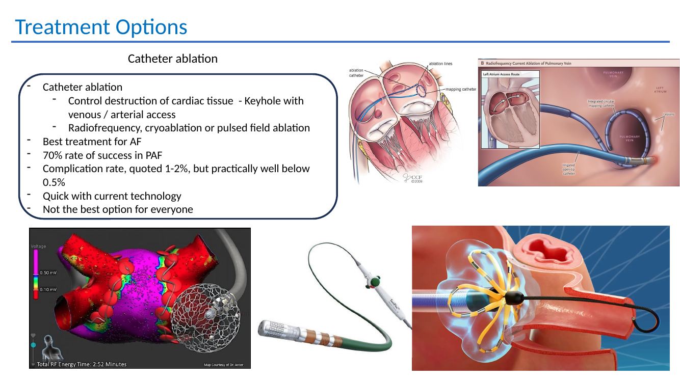
The deck describes ablation as a key treatment for AF, using radiofrequency, cryoablation or pulsed field ablation. For paroxysmal AF, a success rate around 70% is quoted, with contemporary procedural complication rates generally low.

AF catheter ablation slide
AF ablation overview from the original teaching material.
Refer AF / flutter / atrial tachycardiaParticularly useful for symptomatic patients, younger patients, recurrent episodes, medication intolerance or discussion of catheter ablation.
Refer to Dr Handa →

Detailed teaching material

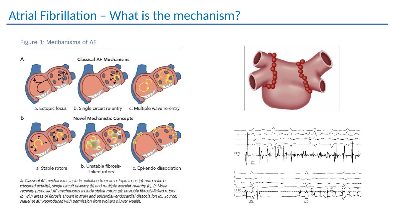
AF mechanism and management framework

The following figures preserve the additional mechanism and management material from the presentation.

Atrial fibrillation symptoms and diagnosis
AF symptoms, diagnosis and additional investigation.
Atrial fibrillation mechanism
Mechanistic framework for atrial fibrillation.
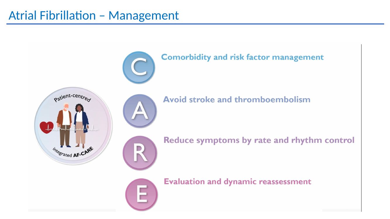
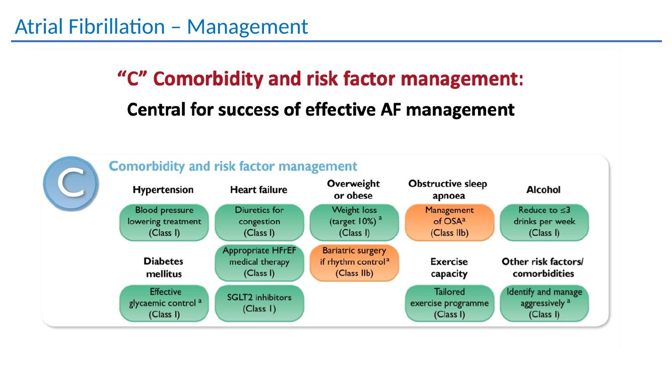
Atrial fibrillation management
AF management teaching material.
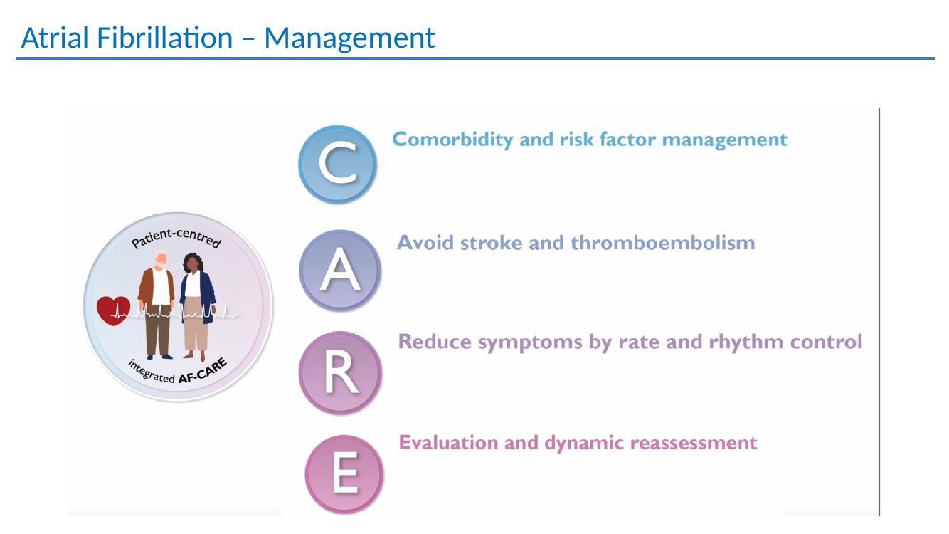
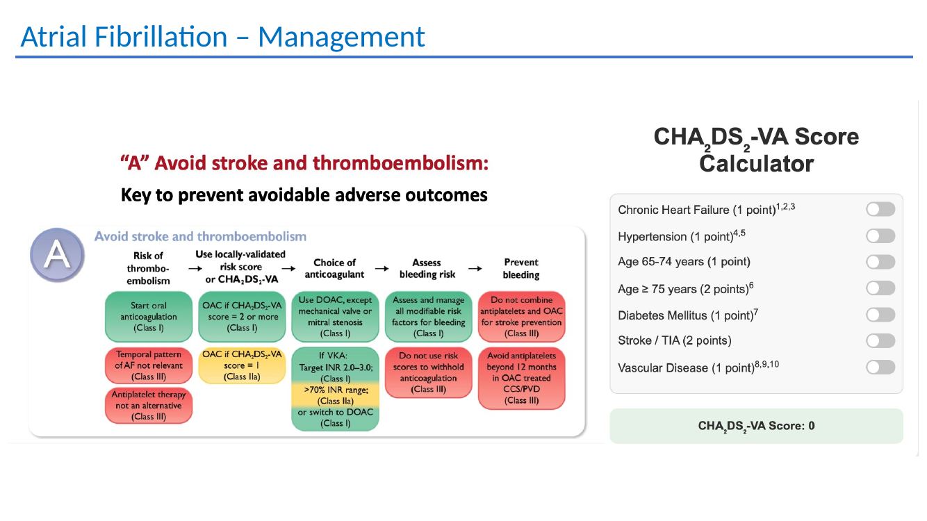
Atrial fibrillation management
AF management teaching material.
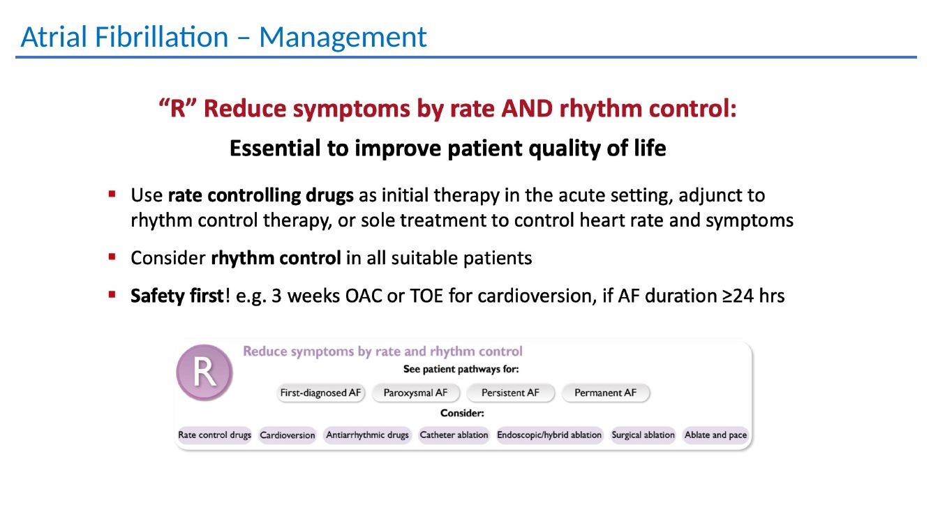
Atrial fibrillation management
AF management teaching material.
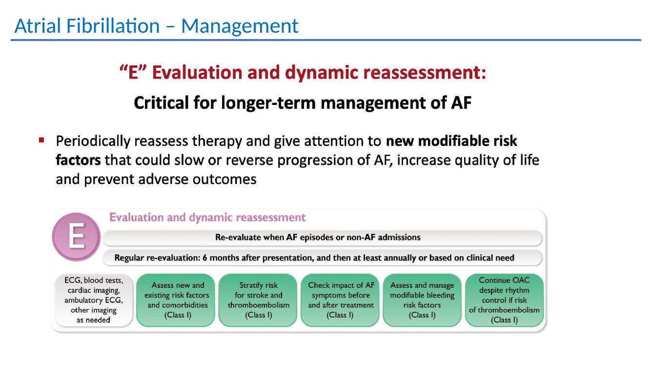
Atrial fibrillation management
AF management teaching material.
Atrial fibrillation management
AF management teaching material.
Atrial fibrillation management
AF management teaching material.
AF treatment options
Drug treatment options and referral considerations.

03 · Atrial flutter & atrial tachycardia

Fast atrial rhythms beyond AF

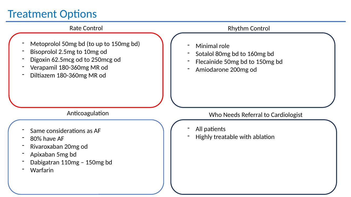
Atrial flutter

Highly amenable to ablation

Rate-control drugs are similar to AF. The presentation notes a limited role for rhythm-control medication and recommends referral of all patients because typical flutter is highly treatable with ablation.

~80%coexistence with AF noted in the deck
Atrial flutter activation mapping from the original teaching presentation.
Atrial tachycardia

Think beyond “sinus tachycardia”

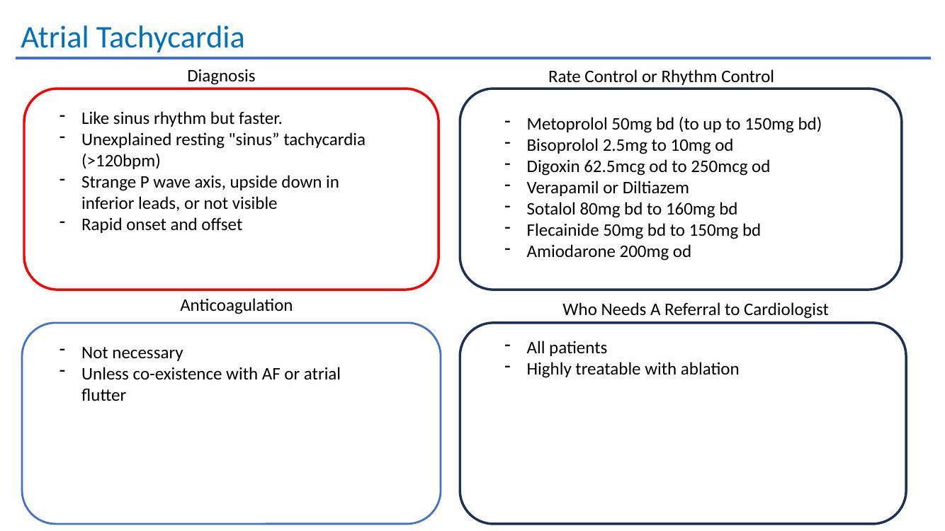
  • Unexplained resting tachycardia >120 bpm
  • Unusual P-wave axis, including inverted inferior P waves
  • P waves may be difficult to see
  • Rapid onset and offset

Anticoagulation is generally not required unless AF/flutter coexist. Rate or rhythm-control medication can be used, but definitive ablation is often effective.

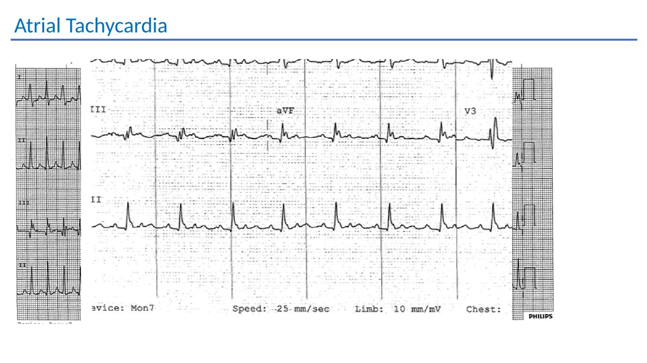
Atrial tachycardia slide
Atrial tachycardia: practical recognition and treatment points.
Atrial tachycardia activation mapping from the original teaching presentation.
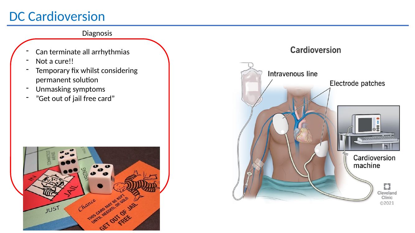
DC cardioversion: can terminate atrial arrhythmias, but it is not a cure. Think of it as a temporary reset while the longer-term strategy is decided.
Refer atrial flutter / atrial tachycardiaTypical flutter and focal atrial tachycardia are often highly amenable to catheter ablation, particularly when recurrent or symptomatic.
Refer to Dr Handa →

Detailed teaching material

Flutter, atrial tachycardia and cardioversion

Atrial flutter treatment options
Rate control, anticoagulation and rhythm-control considerations in atrial flutter.
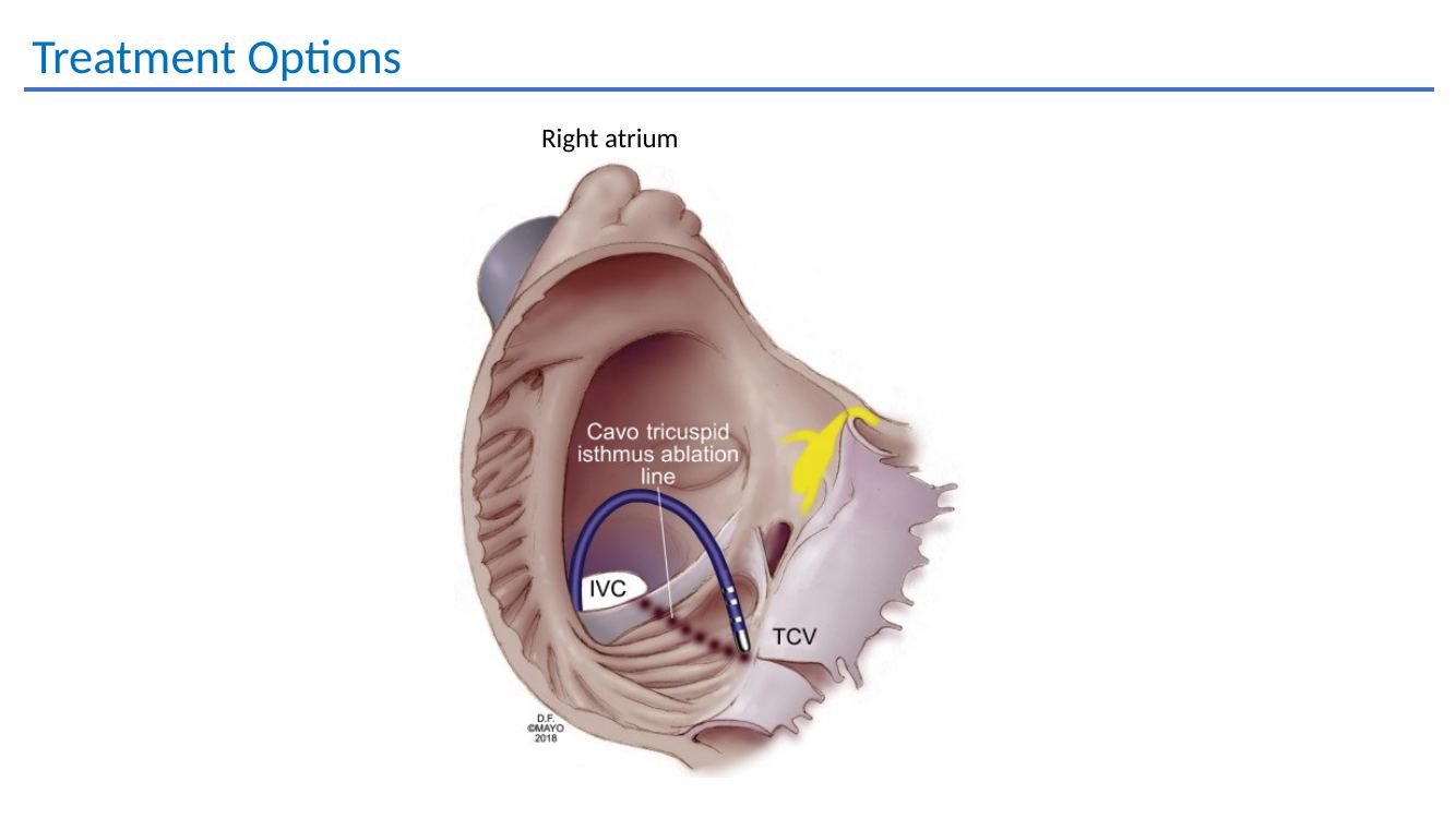
Atrial flutter ablation anatomy
Right atrial anatomy and ablation teaching material.
Atrial tachycardia
Atrial tachycardia teaching example.
Atrial tachycardia diagnosis and treatment
Recognition, pharmacological management and referral of atrial tachycardia.
DC cardioversion
DC cardioversion as a temporary rhythm-restoration strategy rather than definitive cure.

04 · Supraventricular tachycardia

AVNRT and WPW / AVRT

Rapid onset and offset is the recurring clue. Capture the rhythm if possible — a consumer single-lead ECG can be very useful, while a 12-lead ED ECG remains ideal.

AVNRT

Usually irritating rather than dangerous

  • Often random onset, although exercise can trigger episodes
  • Vagal manoeuvres can terminate episodes
  • Beta blockers or non-dihydropyridine calcium-channel blockers may suppress symptoms
  • Anti-arrhythmic drugs have little routine role
  • All confirmed cases are reasonable cardiology referrals
98%+ablation success
1 / 2000quoted AV block / pacemaker risk
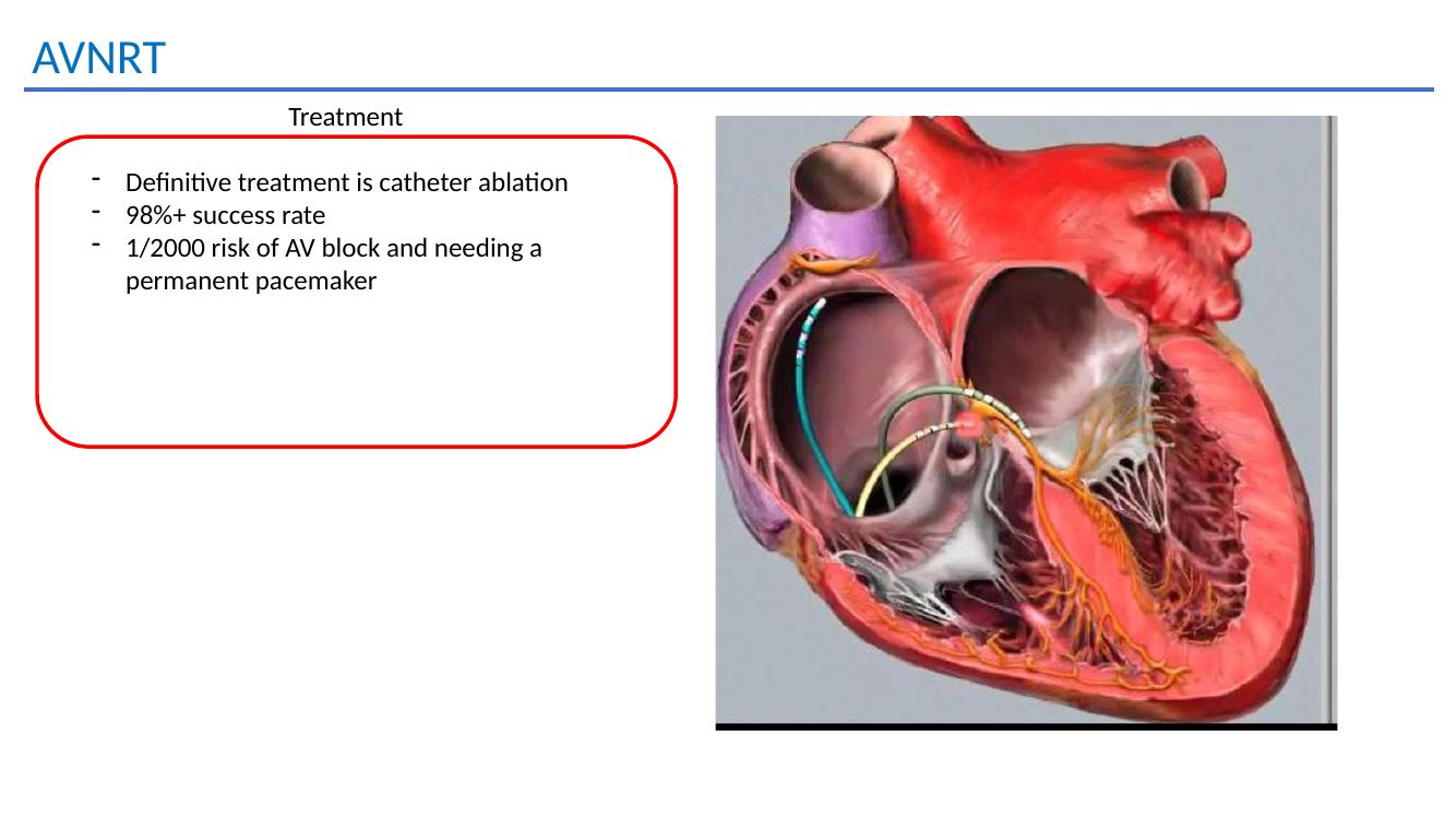
AVNRT treatment slide
Definitive AVNRT treatment is slow-pathway catheter ablation.
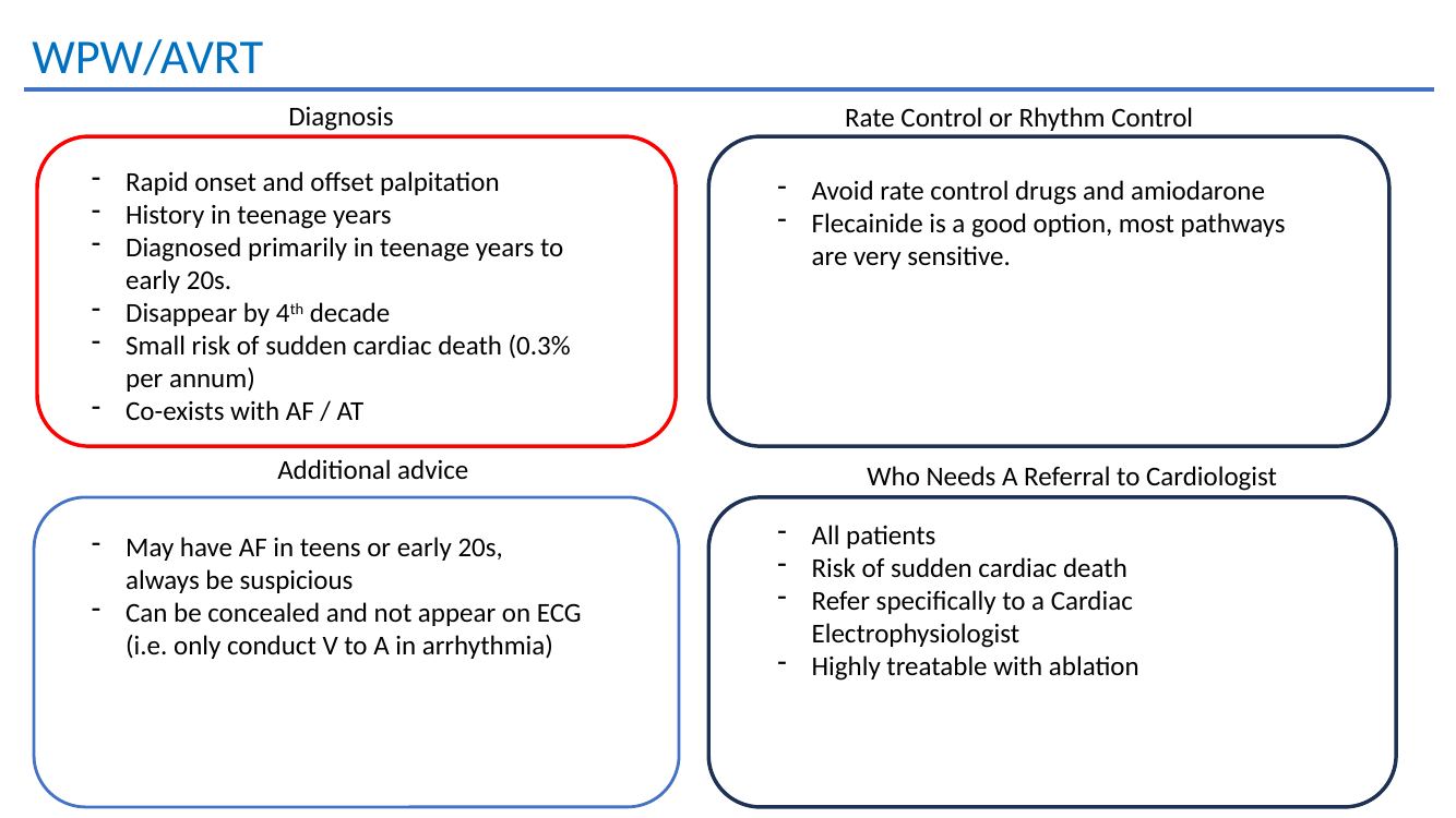
WPW / AVRT

Recognise pre-excitation — and refer

  • Delta waves can be subtle
  • Short PR interval
  • Right-sided pathways may show more obvious pre-excitation
  • Accessory pathways may also be concealed and not visible on resting ECG
  • Often presents in adolescence or early adulthood
  • Flecainide can be effective; avoid AV nodal blockers in pre-excited AF

The deck quotes a small but real sudden-death risk and recommends referral specifically to an electrophysiologist. Ablation is potentially curative.

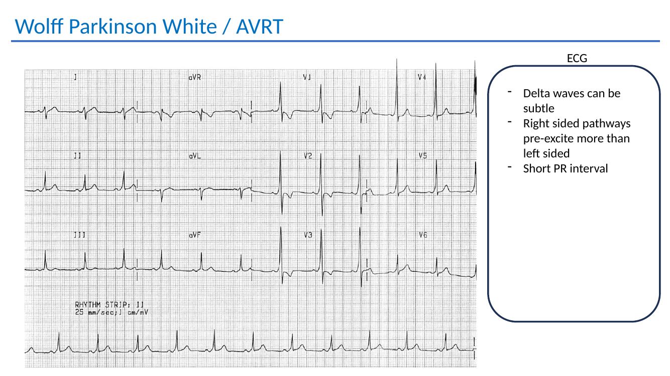
WPW ECG slide
WPW / AVRT ECG teaching: subtle delta waves and short PR can be easy to miss.
Accessory-pathway activation mapping and ablation from the original teaching presentation.
GP practice point: the most useful attachment is often a rhythm tracing. Encourage patients with intermittent episodes to capture a single-lead ECG if available; a 12-lead ECG during tachycardia is ideal. Any WPW / pre-excitation pattern warrants specialist assessment.
Refer SVT / WPWRecurrent abrupt palpitations, documented SVT or ventricular pre-excitation are all appropriate electrophysiology referrals; ablation is often definitive.
Refer to Dr Handa →

Detailed teaching material

AVNRT and AVRT / WPW

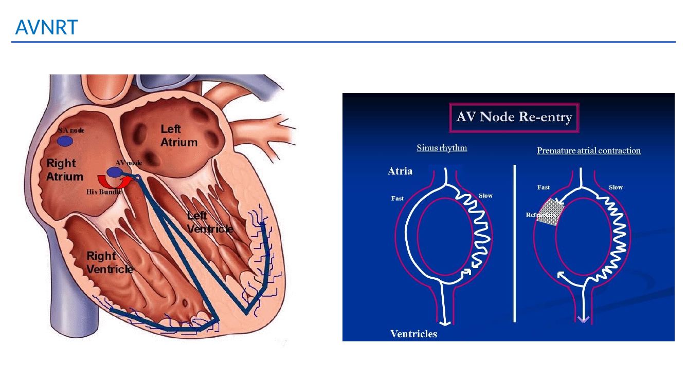
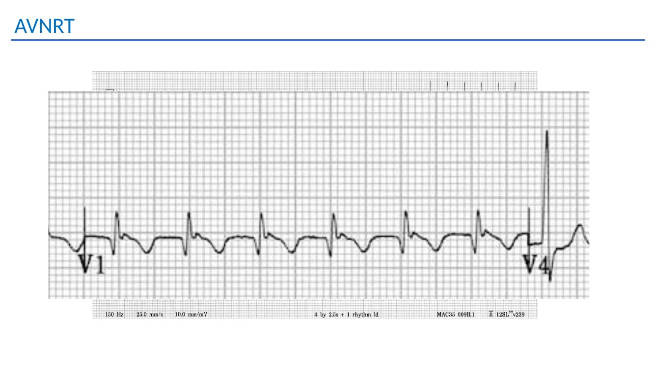
AVNRT teaching slide
AVNRT mechanism and ECG teaching.
AVNRT teaching slide
AVNRT electrophysiology teaching material.
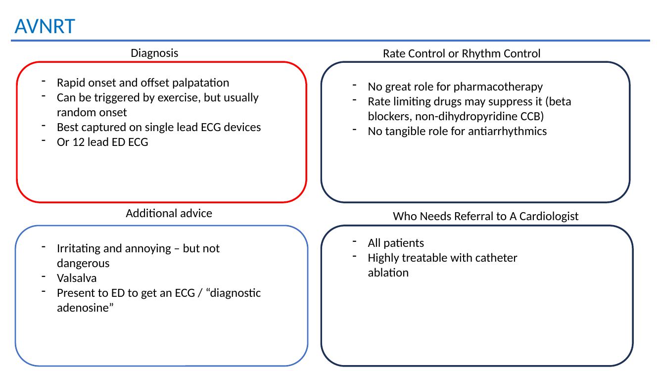
AVNRT practical advice
Practical recognition, monitoring and treatment of AVNRT.
AVNRT catheter ablation
AVNRT catheter ablation, success and AV block risk.
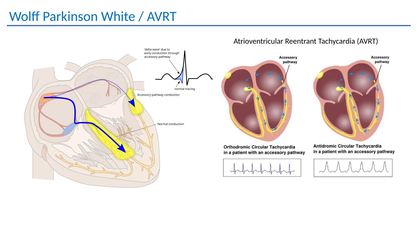
WPW and AVRT mechanism
Accessory-pathway mediated AV re-entrant tachycardia.
WPW practical advice
WPW presentation, sudden-death risk, treatment and referral considerations.

05 · Ventricular arrhythmias

VT, NSVT and ventricular ectopy

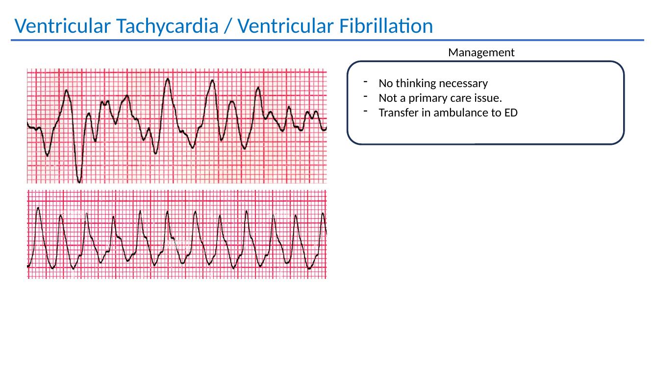
Sustained VT / VFNo primary-care management pathway: transfer by ambulance to an emergency department.

Non-sustained VT

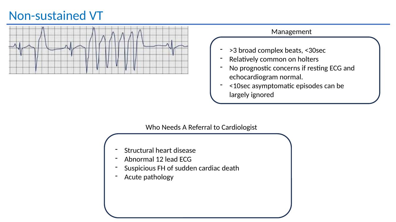
Defined in the presentation as more than 3 broad-complex beats lasting less than 30 seconds. Short asymptomatic episodes can be low risk when the resting ECG and echocardiogram are normal.

Refer when there is

Structural heart diseaseAbnormal 12-lead ECGSuspicious family historyAcute pathology

Ventricular ectopy

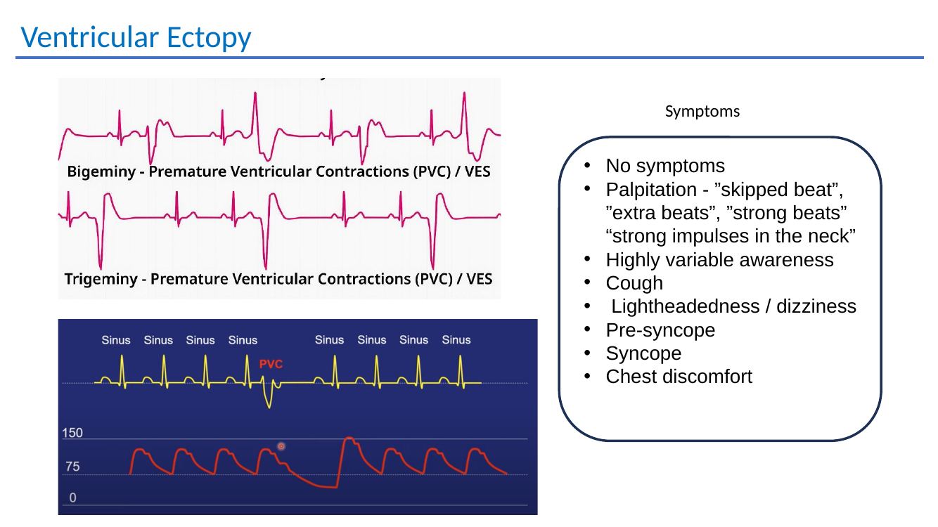
Symptoms range from none at all to skipped beats, forceful beats, cough, dizziness, presyncope, syncope or chest discomfort.

Common contributors

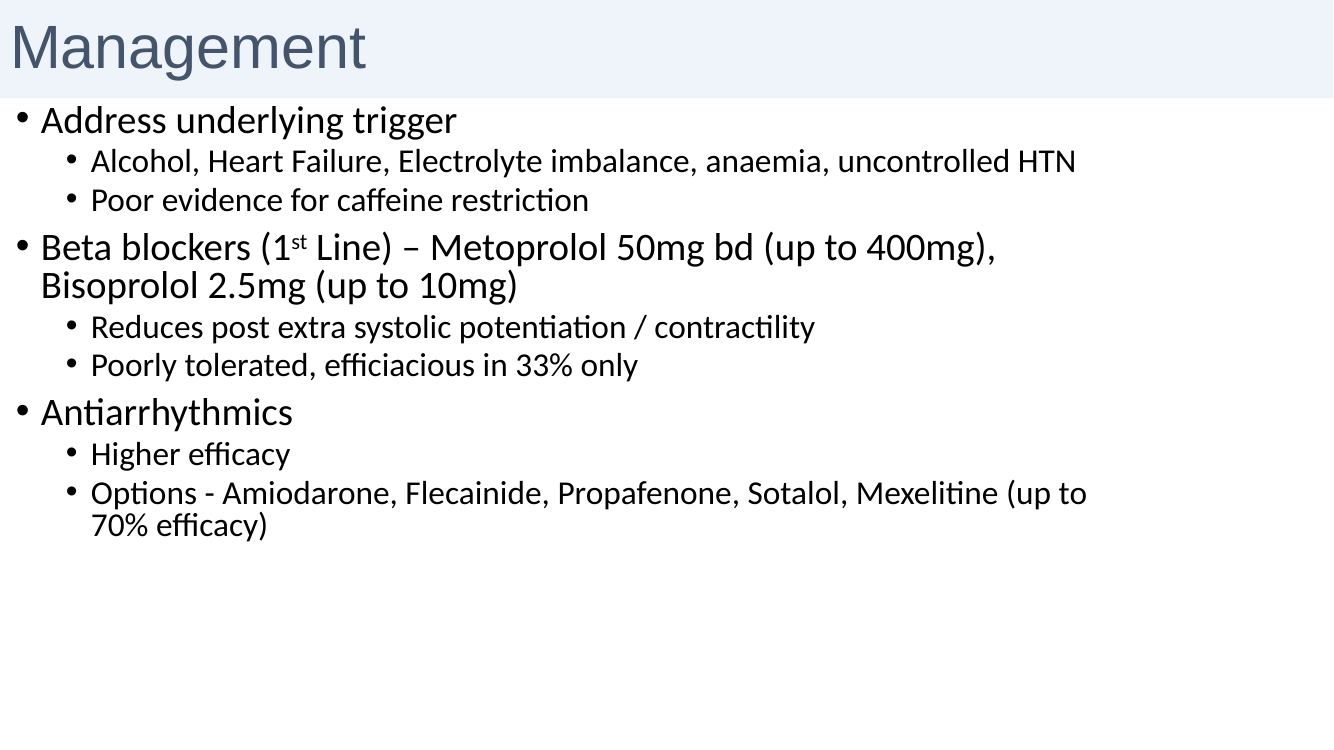
AlcoholHeart failureElectrolytesAnaemiaUncontrolled HTN

The deck notes weak evidence for routine caffeine restriction.

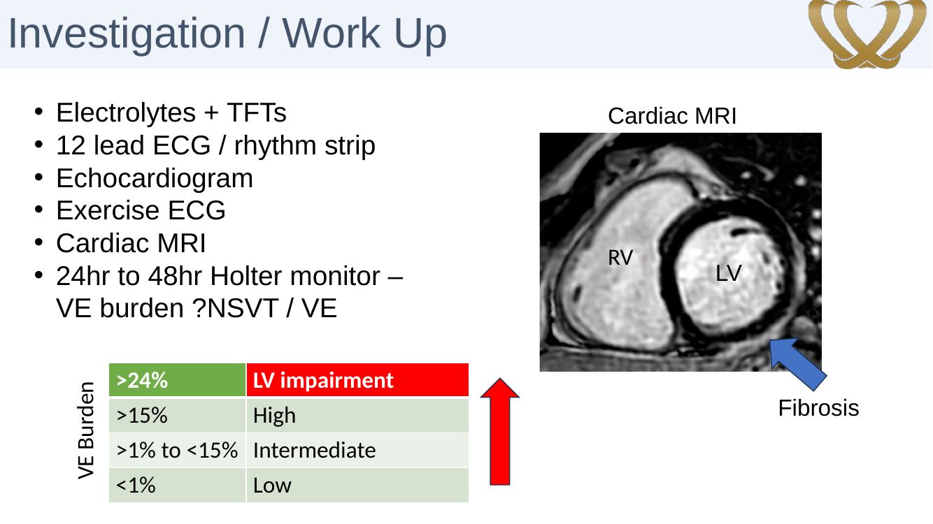
Work-up of ventricular ectopy

  • Electrolytes and TFTs
  • 12-lead ECG / rhythm strip
  • Echocardiogram
  • Exercise ECG
  • Cardiac MRI where appropriate
  • 24–48 hour Holter for burden and NSVT

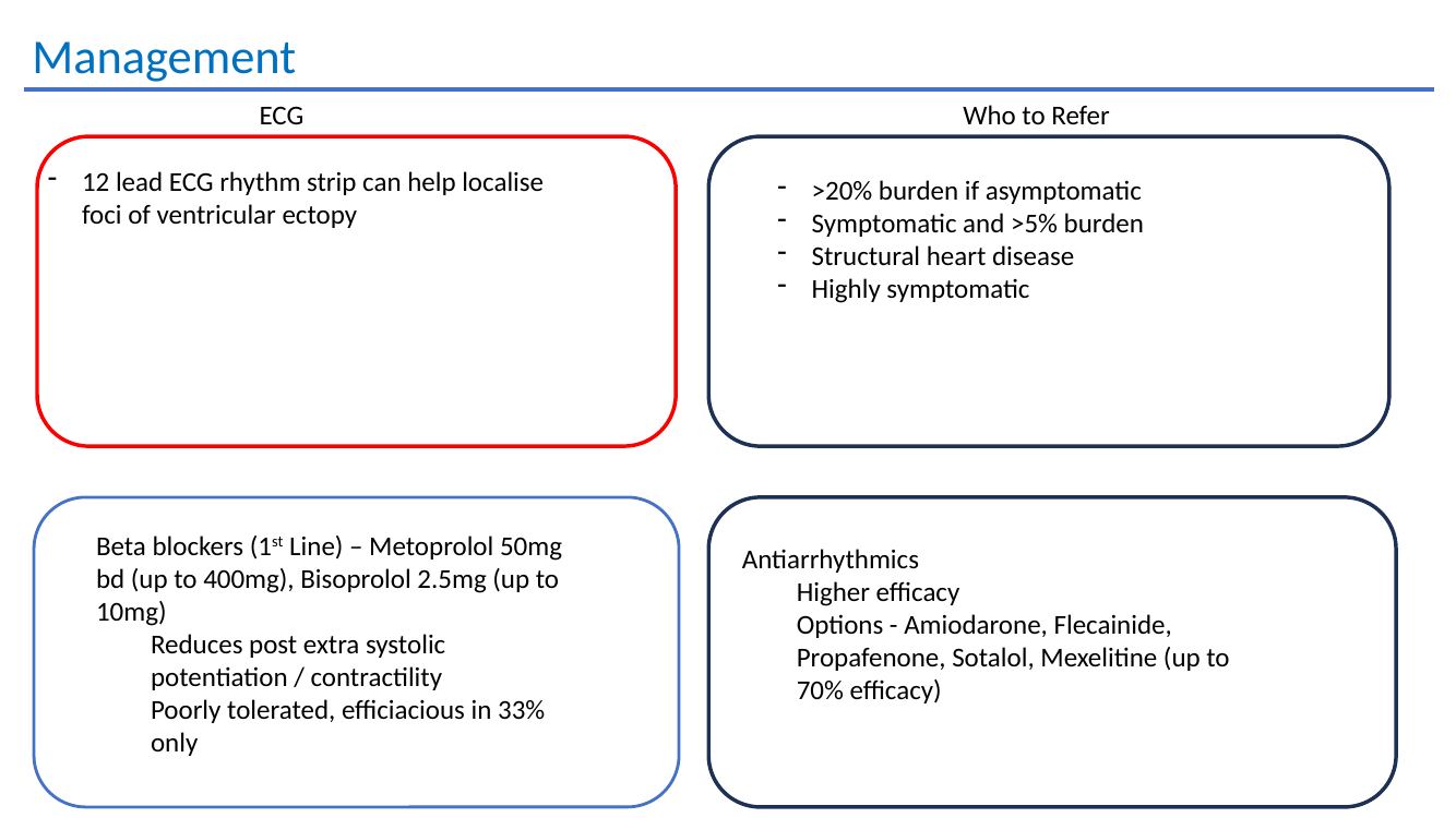
Referral thresholds in the deck

  • >20% burden if asymptomatic
  • Symptomatic and >5% burden
  • Structural heart disease
  • Highly symptomatic patient
Ventricular ectopy burden and cardiac MRI slide
Ventricular ectopy burden and the role of cardiac MRI in assessing myocardial substrate.
Beta blockersFirst line in the presentation; symptom efficacy is modest.
Anti-arrhythmicsFlecainide, propafenone, sotalol, amiodarone or mexiletine can be considered in selected patients.
Catheter ablationPotentially curative and definitive where ectopy is symptomatic or high burden.
12 lead ECG localisation of ventricular ectopy
A 12-lead rhythm strip can help localise the site of origin of ventricular ectopy.
Ventricular ectopy mapping and catheter ablation from the original teaching presentation.
GP practice point: duration alone does not determine the significance of NSVT. The resting ECG, ventricular function, exercise relationship, symptoms, family history and evidence of myocardial scar or cardiomyopathy are what determine how aggressively it should be investigated.
Refer ventricular ectopy / NSVTRefer when symptomatic, high burden, associated with structural disease or an abnormal ECG, exercise-related, or accompanied by syncope or a concerning family history.
Refer to Dr Handa →

Detailed teaching material

Ventricular arrhythmia and ectopy teaching

VT and VF emergency management
Sustained VT / VF: emergency transfer rather than primary-care management.
Non sustained ventricular tachycardia
NSVT definition, low-risk features and referral triggers.
Ventricular ectopy symptoms
Clinical presentation of ventricular ectopy.
Ventricular ectopy management
Triggers, beta blockers and anti-arrhythmic options for ventricular ectopy.

06 · Bradyarrhythmias, pacing & syncope

When slow rhythms need escalation

Symptoms that matter

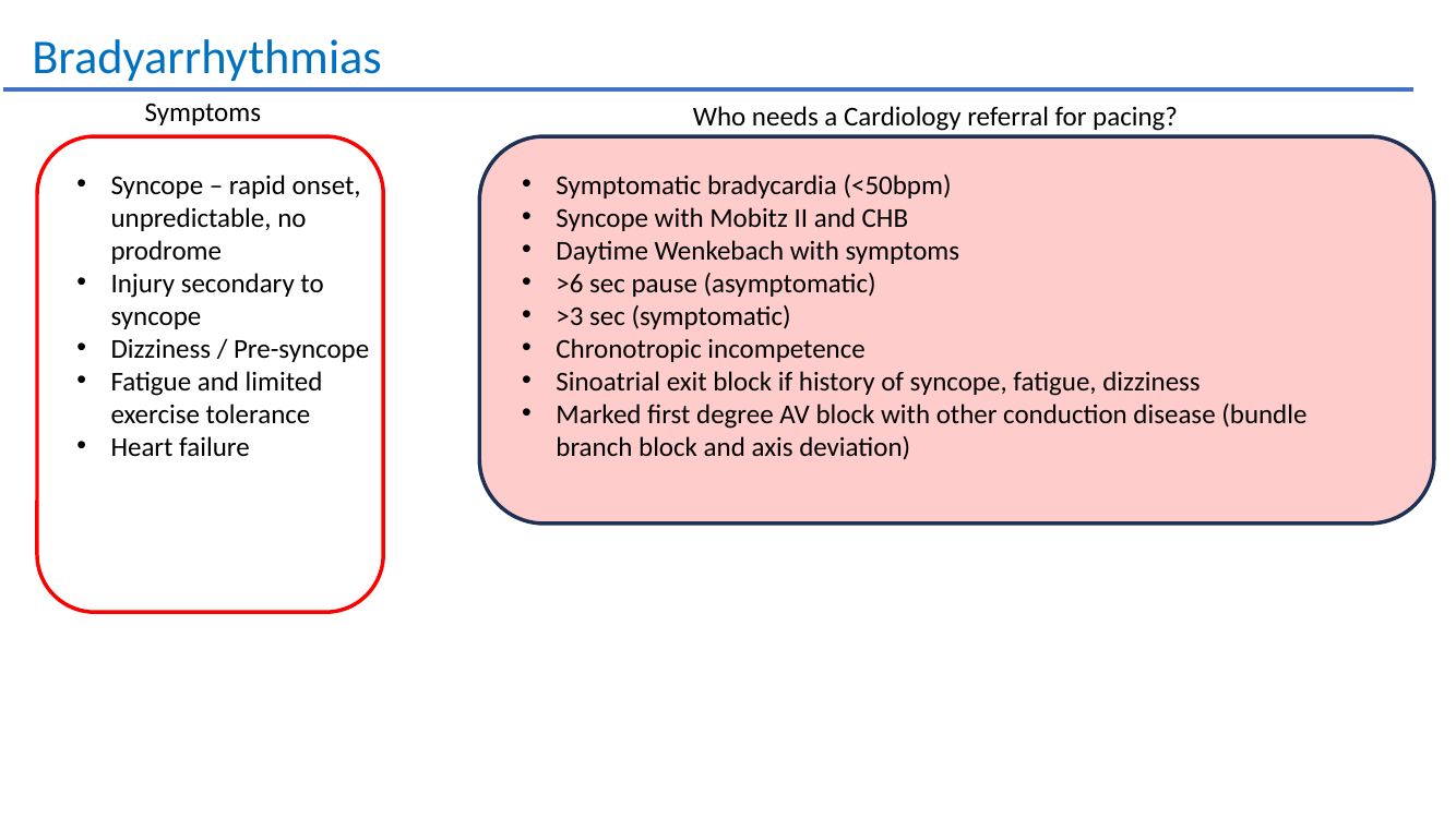
Syncope without warning, injury, dizziness/presyncope, fatigue, reduced exercise tolerance and heart failure.

Pacing signals

Symptomatic bradycardia, Mobitz II or complete heart block with syncope, significant pauses, chronotropic incompetence and advanced conduction disease.

Pacemakers

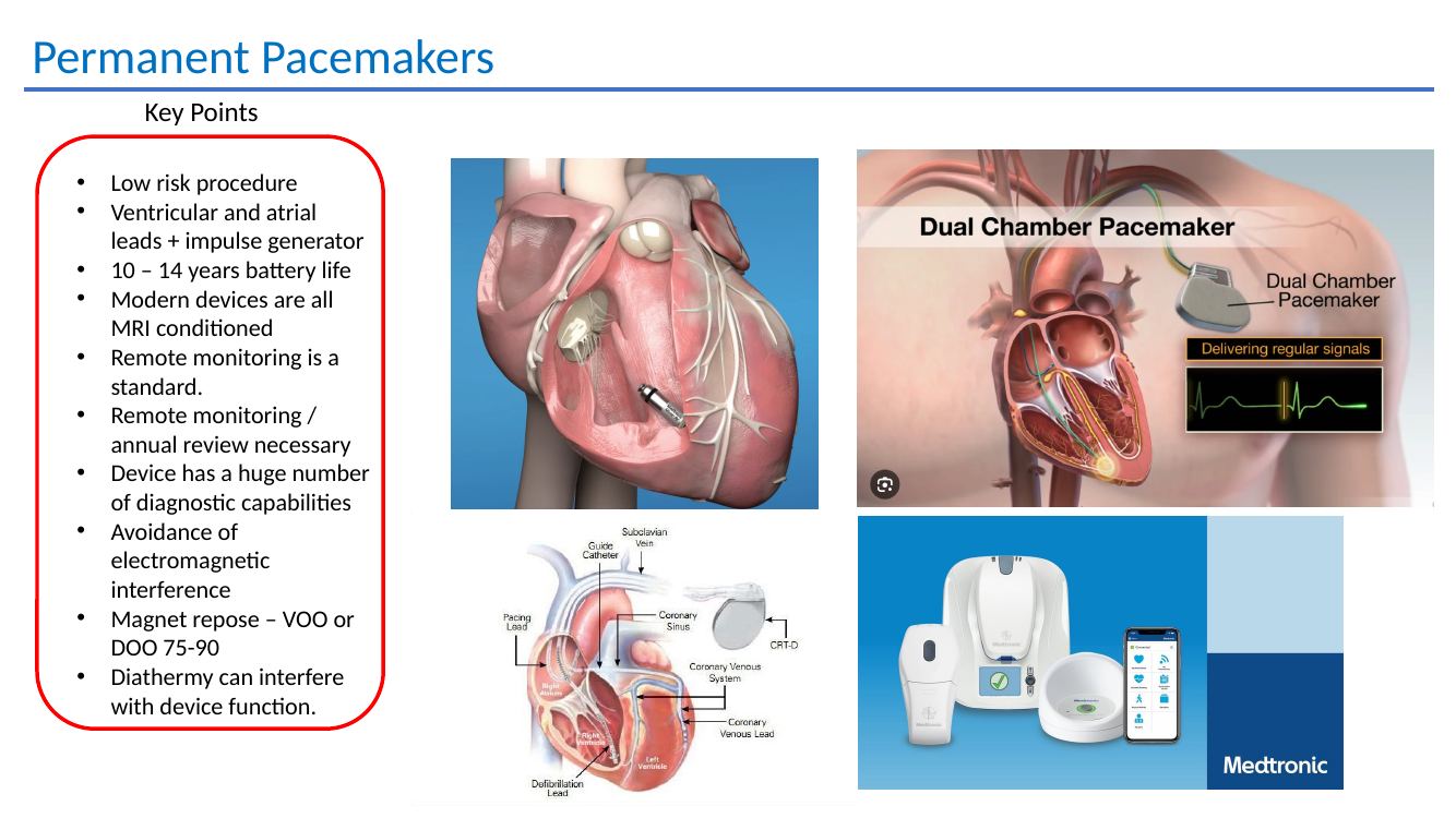
Low-risk implant procedure; atrial and ventricular leads with a pulse generator, typical battery life 10–14 years, MRI-conditional systems and remote monitoring.

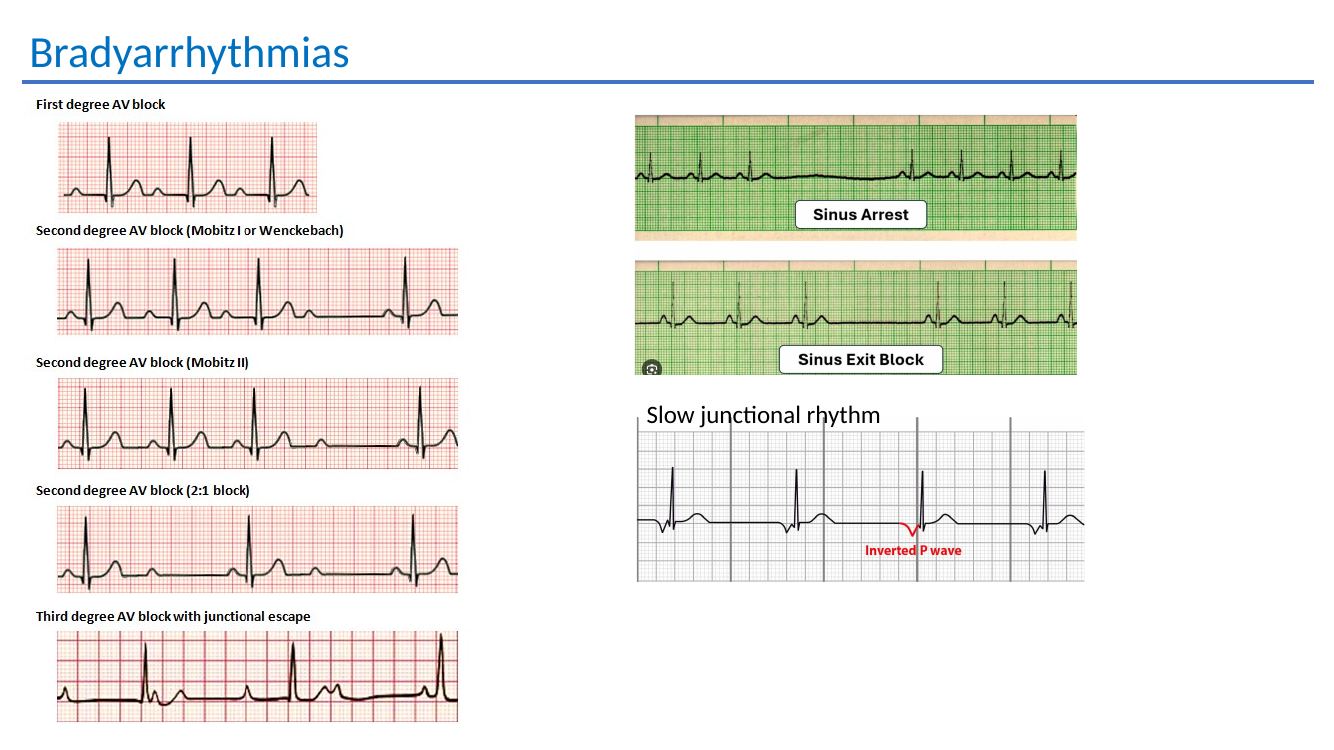
Bradyarrhythmia ECG teaching slide
Bradyarrhythmia ECG patterns from the teaching presentation: first-degree AV block, Wenckebach, Mobitz II, 2:1 block, complete heart block with junctional escape, sinus arrest, sinoatrial exit block and slow junctional rhythm.

Practical referral thresholds

  • Symptomatic bradycardia <50 bpm
  • Syncope with Mobitz II or complete heart block
  • Daytime Wenckebach with symptoms
  • >6 second pause if asymptomatic
  • >3 second pause if symptomatic
  • Chronotropic incompetence
  • SA exit block with syncope, fatigue or dizziness
  • Marked first-degree AV block plus other conduction disease
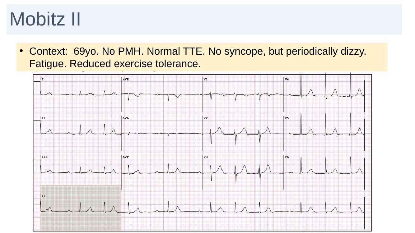
Mobitz II case slide
Case example: Mobitz II in a patient with dizziness, fatigue and reduced exercise tolerance.
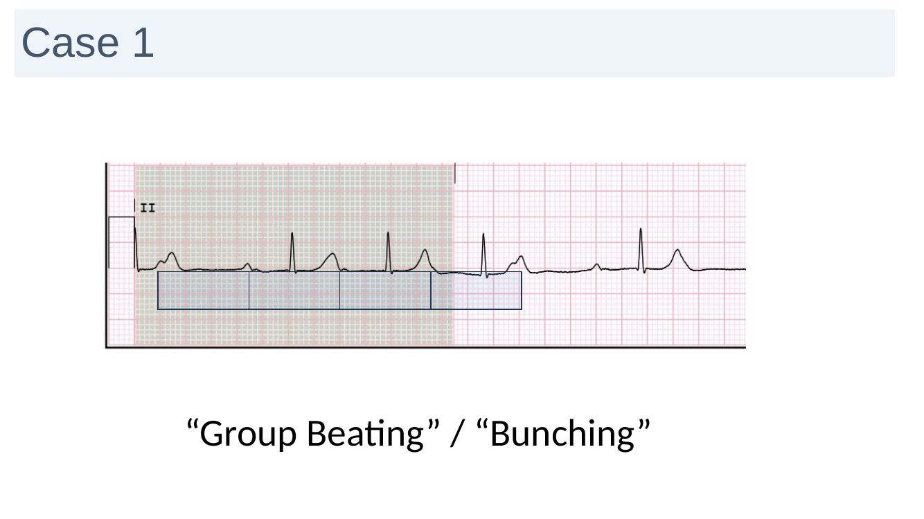
Group beating ECG slide
“Group beating / bunching” — rhythm-strip pattern recognition from the teaching deck.

Implantable loop recorder

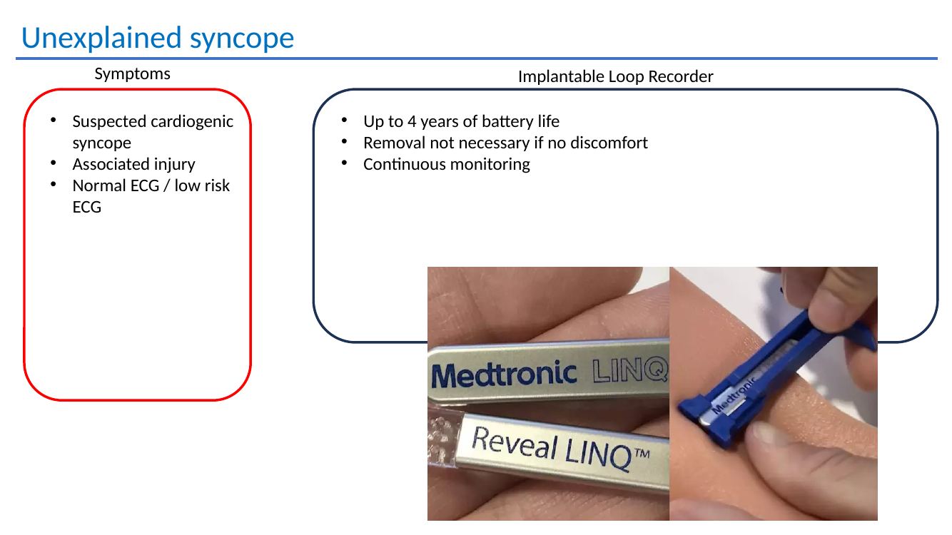
Useful for suspected cardiogenic unexplained syncope, especially where injury has occurred but the baseline ECG is normal or low risk.

  • Continuous monitoring
  • Up to 4 years of battery life
  • Removal is not necessarily required if asymptomatic at end of service
GP practice point: correlate bradycardia or pauses with symptoms and review reversible contributors, but persistent high-grade AV block or symptomatic severe bradycardia is not a routine outpatient problem and should be escalated urgently.
Refer bradycardia / syncopeAppropriate for symptomatic bradycardia, pauses, conduction disease, chronotropic incompetence or unexplained syncope where a cardiac cause remains possible.
Refer to Dr Handa →

Detailed teaching material

Bradyarrhythmias, pacemakers and unexplained syncope

Bradyarrhythmia symptoms and pacing referral
Symptoms and referral thresholds for pacing.
Permanent pacemaker key points
Permanent pacemaker implantation, battery life, MRI conditionality, remote monitoring and electromagnetic considerations.
Implantable loop recorder for unexplained syncope
Implantable loop recorder use in unexplained suspected cardiogenic syncope.

07 · Premature atrial contractions

PACs are usually benign in a structurally normal heart

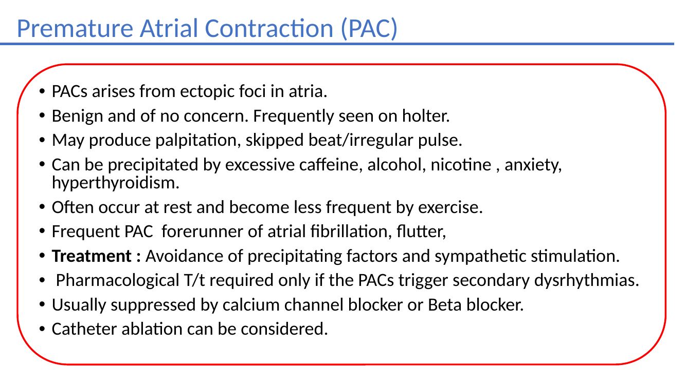
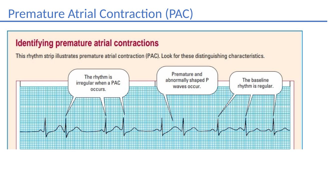
PACs arise from ectopic atrial foci and are frequently seen on ambulatory monitoring. They may cause a skipped beat or irregular pulse and often need only reassurance and trigger modification, although a high burden may be a marker of future atrial arrhythmia.

Common triggers

CaffeineAlcoholNicotineAnxietyHyperthyroidism

They often occur at rest and may become less frequent during exercise. Frequent PACs can be a forerunner of AF or flutter.

Treatment

Avoid precipitating factors and excess sympathetic stimulation. Drug treatment is generally reserved for troublesome symptoms or when PACs trigger another dysrhythmia; beta blockers or calcium-channel blockers may help, and ablation can be considered in selected cases.

Detailed teaching material

Premature atrial contractions

Premature atrial contractions
PAC teaching example.
Premature atrial contractions management
PAC causes, symptoms and management.

08 · Inherited arrhythmia syndromes

Long QT & Brugada

Long QT syndrome

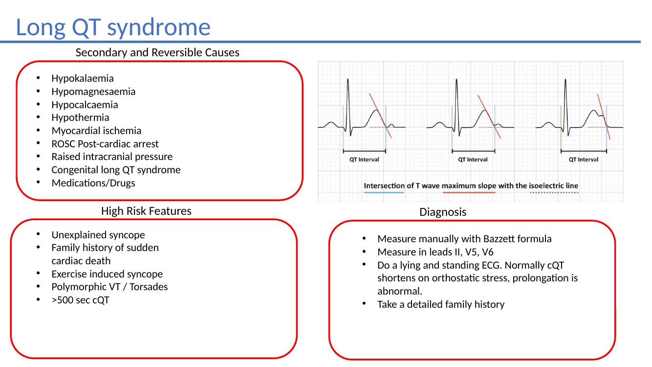
Exclude reversible causes first

HypokalaemiaHypomagnesaemiaHypocalcaemiaHypothermiaIschaemiaPost-arrestRaised ICPDrugs

High-risk features

  • Unexplained syncope
  • Exercise-induced syncope
  • Family history of sudden cardiac death
  • Polymorphic VT / torsades
  • Marked QT prolongation

Practical assessment

  • Measure QT manually, particularly in II, V5 and V6
  • Consider lying and standing ECGs
  • Take a detailed family history
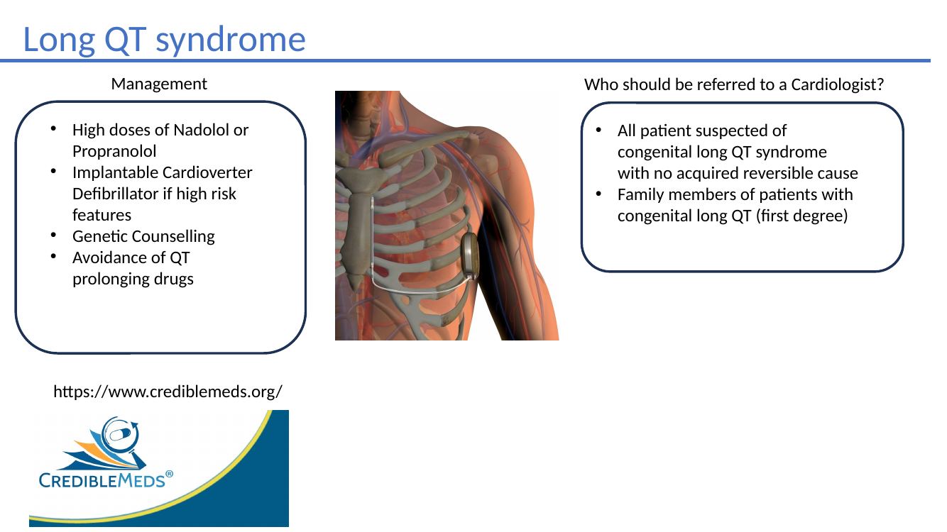
  • Refer all suspected congenital LQTS without a reversible cause
  • Screen first-degree relatives of confirmed congenital LQTS
Long QT syndrome ECG slide
Long QT syndrome ECG teaching example.
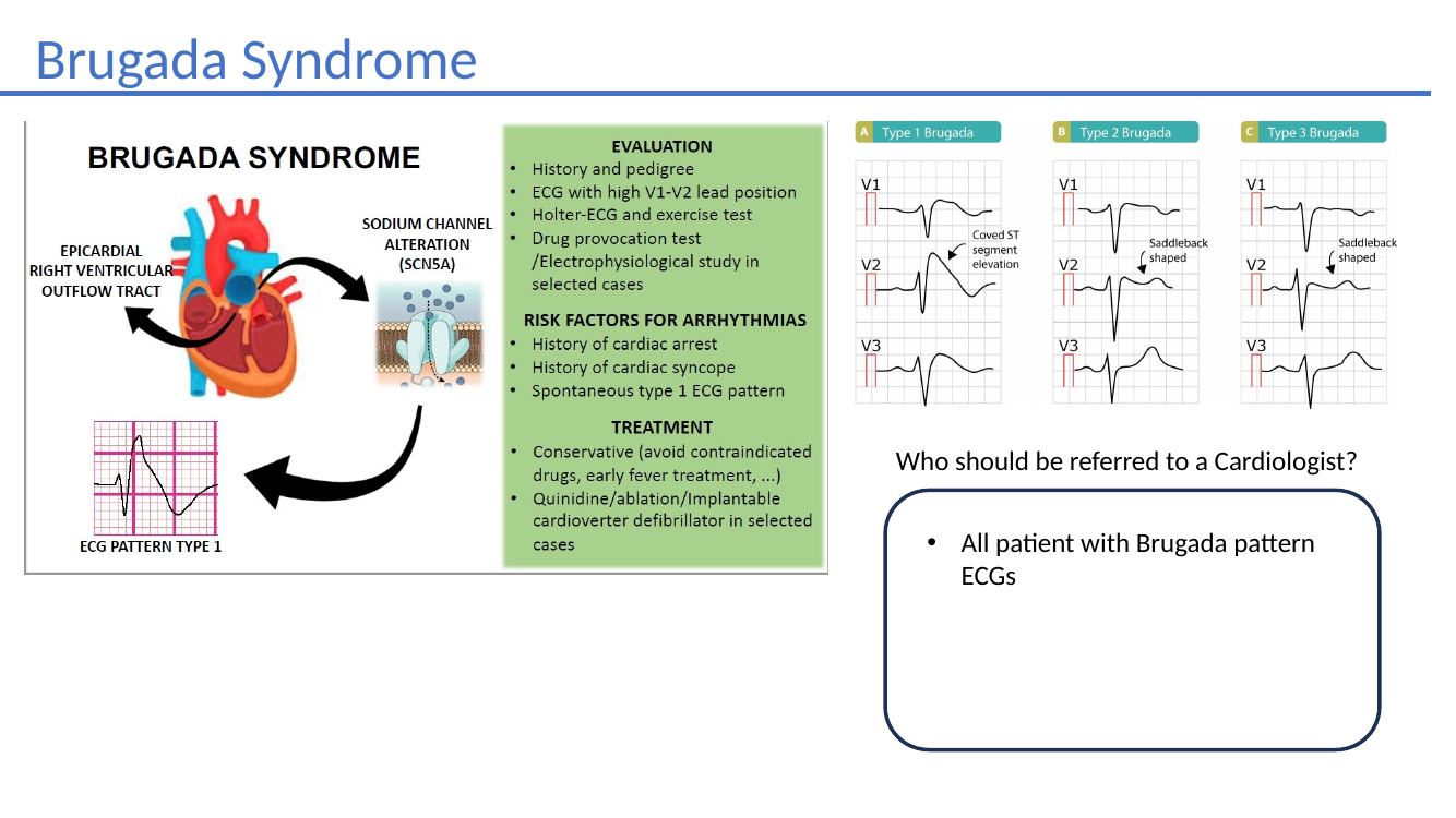
Brugada syndrome

Brugada-pattern ECG = cardiology referral

The deck's rule is deliberately simple: all patients with a Brugada-pattern ECG should be referred for specialist assessment.

Brugada syndrome slide
Brugada syndrome teaching example.
Refer suspected inherited arrhythmiaLong QT, Brugada-pattern ECG, unexplained exertional syncope or a concerning family history of sudden cardiac death warrant specialist assessment.
Refer to Dr Handa →

Detailed teaching material

Long QT and Brugada syndrome

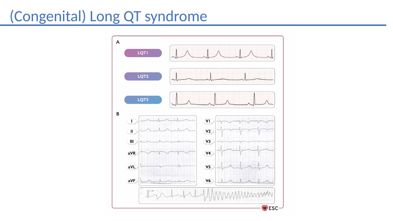
Congenital long QT syndrome
Congenital long QT syndrome teaching material.
Long QT syndrome ECG teaching
Long QT syndrome ECG examples.
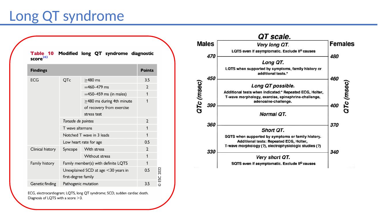
Long QT syndrome management
Long QT management, beta blockade, ICD consideration, genetic counselling and referral.

Clinical resources

Guidelines behind the practical approach

The core of this website comes from Dr Handa’s GP teaching material. These external resources provide the broader Australian and international guideline context for clinical decision-making.

Clinical resources reviewed September 2026. Always use current product information, local hospital pathways and individual clinical judgement.

Dr Balvinder Handa
Dr Balvinder Handa MBBS BSc MRCP FRACP PhD

About Dr Handa

Cardiac electrophysiology, devices and general cardiology

Dr Balvinder Handa is a Consultant Cardiologist and Cardiac Electrophysiologist with expertise in the diagnosis and management of heart rhythm disorders and cardiac devices. His practice combines advanced electrophysiology with comprehensive general cardiology care.

His areas of expertise include catheter ablation of arrhythmias, including atrial fibrillation; implantation of pacemakers, implantable cardioverter defibrillators and cardiac resynchronisation therapy devices; lead extraction; complex vascular access and venoplasty; and the diagnosis and management of inherited cardiac conditions. He also has extensive experience in echocardiography.

Dr Handa completed his undergraduate medical training and PhD at Imperial College London, followed by general cardiology training and a 3-year fellowship in Cardiac Electrophysiology and Devices at Hammersmith Hospital, Imperial College Healthcare NHS Trust. After qualifying as a consultant in the UK, he relocated to Perth and now works across both public and private practice, including a public appointment at Sir Charles Gairdner Hospital and private practice at Hollywood Private Hospital.

He is actively involved in medical education and research, including peer-reviewed publications, book chapters, a registered clinical patent and international research awards. He is also a Clinical Lecturer / Clinical Tutor at the University of Notre Dame Medical School.

Catheter ablationAF & SVTPacemakers & ICDsCRTInherited cardiac conditionsGeneral cardiology

Referral & contact

Dr Balvinder Handa

Consultant Cardiologist & Cardiac Electrophysiologist

Special interests include cardiac arrhythmias, catheter ablation, cardiac devices, inherited cardiac conditions, general cardiology, ECG and Holter reporting.

General Cardiology Training & PhD — Imperial College London3-year Electrophysiology & Devices Fellowship — Imperial College London & Sir Charles Gairdner HospitalPublic Appointment — Sir Charles Gairdner Hospital
Perth Cardiovascular Institute logo

Perth Cardiovascular Institute

Refer to Dr Balvinder Handa

Perth Mount Medical Centre
Suite 12, 1st floor
146 Mounts Bay Road, Perth
This website is educational material for health professionals. It is intended as a practical reference and does not replace individual clinical assessment, current prescribing information, emergency care pathways, or local hospital protocols.

Refer a patient